Call Us on0510-85386636


Contact: Manager Yang
Hotline: 950-4048-3964 (free)
Tel: 0510-85386636
Mobile: 18011518665
Shangmeng Technology Wuxi Co., Ltd.
Address: A1-602, Tianan Smart City, No. 228 Linghu Avenue, Xinwu District, Wuxi City, Jiangsu Province

[1]Science: Scientists successfully analyze the key structure of HIV virus to overcome major problems
DOI: 10.1126/science.aah5163
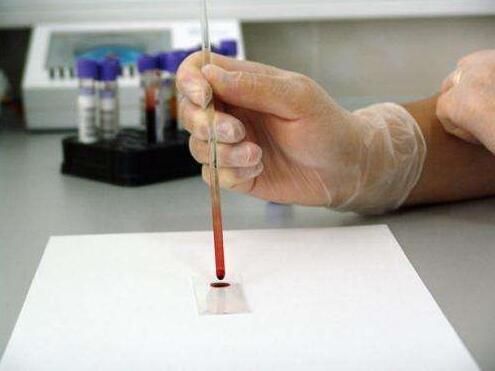
Scientists at the Salk Institute in the United States have recently analyzed the atomic structure of a key part of the HIV virus. This key structure, called the intasome, helps HIV integrate into human host DNA and replicate in the body. The findings are published in the international academic journal Science, which helps develop new HIV treatments.
The author Dmitry Lyumkis said: "HIV is a very intelligent virus, learn how to escape the best drugs. Deep understanding of the virus escape mechanism, the development of more applicable drugs will be a major direction of future research."
In this new study, the researchers used a single-particle cryo-electron microscope that allowed scientists to capture images from larger, complex dynamic molecules. They added a special protein to the viral integrant to promote the solubility of the integrant in glycerol, and added some salt ions to prevent protein aggregation.
[2] Science: How does sleep improve the memory of knowledge?
DOI: 10.1126/science.aah5982
We all know that if you want to consolidate the knowledge you learn during the day, it is best to have a good night's sleep. Although scientists have long known that our memories are stored in the brain's neuronal connections, it is unclear how sleep plays a role in the storage and consolidation of information.
Today, there are two studies that can help us explain this long-standing puzzle.
In fact, we are all curious as to why we must have a certain amount of sleep every day. The latest hypothesis is that sleep can help our brains discharge toxic proteins produced during the day. Recent studies have shown that if we do not get enough sleep, our risk of cardiovascular disease and type 2 diabetes will be significantly improved, not to mention Parkinson's and other neurodegenerative diseases.
Now, researchers from the University of Wisconsin and Johns Hopkins University offer another benefit of sleep, which is the ability to fine-tune the memories of our day-to-day learning. This so-called "synaptic steady state regulation theory" is not the latest product. Researchers from the University of Wisconsin raised the hypothesis 10 years ago that they believe that the connections between brain neurons in the sleep phase will be disconnected, making memory clearer.

[3]Science: The brain is 100 times more complicated than we think.
News reading: Brain is 10 times more active than previously measured, bank find
A recent study by scientists at UCLA may be able to change our previous perception of the human brain. This change may have an important impact on the development of neurological disorders and artificial intelligence.
The focus of this study is on the domain function of brain axons. A neuron is a bulky tree-like structure composed of a cell body and several synapses that grow above it. Previously, researchers have always believed that the signal of neurons is first emitted by the cell body, and the axons simply transfer the above signals passively to other neurons. But this hypothesis has not really been tested. This process is also closely related to the generation and storage of memory.
However, UCLA researchers have found that axons of neurons are not just functions that passively transmit signals. Their research shows that during the movement of animals, the axons can be actively activated, and the relative cell bodies can produce more than 10 times of dendrites. This finding challenges the existing view that the branching of the cell body is the foundation of understanding, memory, and brain activity during learning.
[4] Science: A new breakthrough in cancer immunotherapy! PD-1-activated T cells also need to rely on CD28 co-stimulation
Doi:10.1126/science.aaf0683
Anticancer drugs that block the PD-1 pathway (also known as immunological checkpoint inhibitors) are now approved by the US Food and Drug Administration (FDA) for the treatment of melanoma, lung cancer and several other cancers. These drugs are often described as "releasing the brakes" on the surface of T cells that are dysfunctional.
In a new study, researchers from the Emory Center for Vaccines at Emory University School of Medicine and the Winship Cancer Institute confirmed that even if the brakes imposed on PD-1 are released, these tumors Specific T cells still require "fuel" for proliferation and recovery of an effective immune response. This fuel comes from co-stimulation based on CD28 molecules. The relevant research results were published online in the Science Journal on March 9, 2017, and the title of the paper is "Rescue of exhausted CD8 T cells by PD-1 - targeted therapies is CD28-dependent".
Despite the success of PD-1 targeted drugs, many patients' tumors do not respond to them. The findings of this study suggest that CD28 present on the surface of T cells may be a clinical biomarker that predicts whether PD-1 targeted drugs are effective. In addition, the need for CD28 suggests that co-stimulation may be lost in some patients, which may help guide the design of combination therapies.

[5] Science: What? Can it be infected?
DOI: 10.1126/science.aak9748
Similar to yawning, scratching is also a contagious behavior. Just seeing others scratching their heads can trigger our desire to tickle.
Recently, researchers have found that mice may have the same response, and this finding may help us find a brain circuit that can feel the irritating feeling of others.
Previous research on infectious behavior has been controversial. There is no unified explanation for whether we will behave similarly when we see others scratching or yawning. Therefore, the cause of this contagious behavior is not clear.
Recently, researchers have found that mice have the same feelings as humans, that is, they can also share scratching behavior with their peers.
First, the researchers locked a pair of mice in a cage and analyzed their behavior. The results showed that if one mouse began to scratch, the chance of scratching the other mouse would also increase significantly. To verify that this behavior occurred only because of the visual perception, the researchers allowed the mice to watch the tickling behavior of the mice in the video and obtained similar results.
[6]Science: Two-thirds of carcinogenic mutations are attributed to random DNA replication errors
Doi:10.1126/science.aaf9011 doi:10.1126/science.aam9746
In a new study, researchers from the Johns Hopkins University Kimmel Cancer Center provided evidence that random unpredictable DNA replication "errors" caused nearly two-thirds of cancer-causing mutations. Their research is based on a new mathematical model developed from DNA sequencing data and epidemiological data from around the world. The results of the study were published in the March 24, 2017 issue of Science, titled "Stem cell divisions, somatic mutations, cancer etiology, and cancer prevention."
Cristian Tomasetti, Ph.D., co-author of the paper and assistant professor of biostatistics at the Johns Hopkins University's Kimmel Cancer Center, said, "It is well known that we must avoid environmental factors such as smoking in order to reduce our risk of cancer. But not What is known to many people is that every time a division breaks, a normal cell will replicate its DNA and produce two new daughter cells. There are many mistakes in this process. These replication errors are an important source of carcinogenic mutations. In the past, these oncogenic mutations were scientifically underestimated. This new study is the first to estimate the proportion of mutations caused by these replication errors."

[7] Science: Why do everyone look different? See how scientists explain
DOI: 10.1126/science.aal2913
Although the genes that control the formation of faces in each person are roughly the same, each face is unique. Filippo Rijli and his team discovered an epigenetic mechanism that regulates facial morphogenesis. During early development, neural crest cells that form different facial structures are able to maintain chromosome plasticity, and all genes involved are in a ready state to respond to local signals. Once the cells are exposed to environmental signals, the genes of the neural crest cells change from a ready state to an active state, inducing a position-specific transcription process to form structures such as the chin, the tibia, and the forehead.
Neural crest cells form most of the skull, facial cartilage, and bone. During the early embryonic development, neural crest cells migrate from the developing neural tube to the future head region. These migrating neural crest cells are natural pluripotent cells that, once they reach their final destination, will have fate decisions that differentiate toward the cartilage.
The neural crest cells also acquire a specific location identity to determine the shape of the bones and cartilage, which will form the mandible and chin, the tibia, the nose, and the forehead in the future. The acquisition of such locational identity by cells during migration depends on the path of their migration and their interaction with the local environment. But even if the positional identity is irreversibly determined after migration, the neural crest cells will still maintain some plasticity.
[8]Science: Scientists develop new genome-wide amplification methods that are significantly more efficient than others
DOI: 10.1126/science.aak9787
Recently, in a research report published in the international magazine Science, researchers from Harvard University have developed a new type of whole genome amplification method, which is superior to other genomic amplification methods currently used; In this study, the researchers described the technique and illustrated how it can be used to determine single nucleotide changes in human cells after exposure to UV radiation.
As scientists continue to fully understand the body's genome, new research tools are also being born. One of the researches is to explore the differences between human cells, such as embryonic cells. Each cell has its own uniqueness. The genome, even in the same organism; in the previous study, the researchers developed tools that amplify the differences between cells, which not only help to better understand the principles of genome work, but also have some practical Applications; researchers have developed a tool called MALBAC to study and measure genetic changes between single cells that can be used to screen embryos in in vitro fertilization, but researchers have pointed out that this technique is often limited. Loss of alleles, which often limits their understanding of the process of single nucleotide mutations.
[9] Science: Overturning conventional cognition! Scientists have discovered that dendritic cells may be derived from special progenitor cells
DOI: 10.1126/science.aag3009
Dendritic cells are the "guards" of the body's immunity, which can help the body to effectively detect and activate immunity against foreign pathogens or foreign bodies. So far, researchers believe that the subtypes of dendritic cells are from a common ancestor. Cells have been differentiated; recently, a study published in the international journal Science, researchers from institutions such as the Singapore A*STAR Institute found that the immune cells of the human body may be derived from specific progenitor cells. The research has provided new clues for the later development of new vaccines and the optimization of vaccines.
Professor Andreas Schlitzer, a researcher at the University of Poon, said that our blood is not just red blood cells. Red blood cells are very important for transporting oxygen. The blood also contains many types of immune cells, which can help protect against external pathogens, such as viruses. Or bacteria, etc., researchers have long analyzed the blood immune cell compartment, in which human dendritic cells are the "important interface" between the innate immune system and the immune system adaptive branch. Therefore, the relevant research results are of great significance for understanding the key role played by immune cell subtypes during the regulation of human immune response.
[10] Science is heavy! Scientists have discovered five new blood immune cells!
DOI: 10.1126/science.aah4573
Scientists have discovered several new immune cells in the human immune system.
These cells are a new subpopulation of blood leukocytes called dendritic cells and monocytes. The researchers discovered two new dendritic cell subsets and two new monocyte subsets. They also discovered a new dendritic cell precursor cell, and related research results have recently been published in Science.
Researchers from Broad and other institutions used a technique called single-cell genomics to analyze gene expression patterns in human blood cells. Previously, different immune cells have been studied and classified according to the proteins on their surface. This new technology is more powerful and can reveal rare cell types that were not found in old technology.
The surface of dendritic cells presents a molecule called an antigen. These molecules are recognized by T cells, which then initiate an immune response. Monocytes, the largest blood white blood cells, develop into macrophages responsible for digesting cell debris.
[11]Science: The cause of anti-HIV drug failure may be due to certain vaginal bacteria
Doi:10.1126/science.aai9383 doi:10.1126/science.aan6103
Around the world, the HIV virus infects more than 1 million women every year. In a new study, researchers from Canada, the United States, South Africa, and Sweden found that some types of vaginal bacteria may interfere with drug gels designed to prevent the risk of HIV infection. The results of the study were published in the June 2, 2017 issue of Science, entitled "Vaginal bacteria modify HIV tenofovir microbicide efficacy in African women."
These findings were based on a 2010 study of a South African woman using tenofovir, a microbicidal drug in the form of a vaginal gel, to assess how well it can prevent HIV transmission.
The drug has been shown to be successful in preventing high-risk men from contracting HIV, but the results of women's studies are “disappointing”.
A randomized clinical trial called CAPRISA 004, conducted in 2010, has demonstrated that tenofovir gels reduce HIV infection by 39% before and after sexual contact.
During the study, the researchers studied a subset of women who were still infected with HIV despite the frequent use of this gel.
[12] Science: Discovering a new mechanism of cancer cell migration
Doi:10.1126/science.aal4713
In a new study, young researchers from France discovered a new mechanism to promote cell migration. The cell produces a variety of small hooks on its cell membrane surface that help it attach to the extracellular collagen fibers and migrate along these collagen fibers. This effect helps us better understand that cells escape from the tumor mass and migrate and form new lesions in the body. The results of the study were published in the June 16, 2017 issue of Science, entitled "Tubular clathrin/AP-2 lattices pinch collagen fibers to support 3D cell migration". The author of the paper is Guillaume Montagnac, research leader of the French National Institute of Health and Medical Research, and Nadia Elkhatib, researcher of the Costa Rica Husi Institute of the University of Paris-Sacré, France.
Cell migration is a normal process that is vital to life. In oncology, it is involved in new metastases.
Guillaume Montagnac claims, “Before, we knew that cells depend on certain structures to attach themselves to its environment. We have now identified a new called clathrin-coated pit. Structures, which are known to play an important role in other cellular functions. Cancer cells use them as hooks to attach to other structures to move around. These new structures cause about 50% of cells to attach to the surrounding structure."
[13]Science: High-throughput analysis of thousands of micro proteins, is expected to trigger protein engineering changes
Doi:10.1126/science.aan0693 doi:10.1126/science.aan6864
Advances in DNA synthesis technology combined with improvements in the use of computational methods to design new proteins are preparing for a new era of data-driven protein molecular engineering.
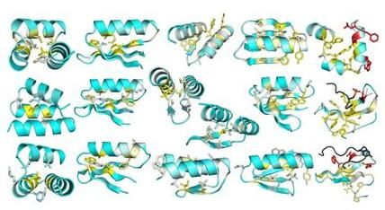
In a new study, researchers from the University of Washington and the University of Toronto in Canada reported a new high-throughput method that makes the folding stability of computationally designed proteins (designed proteins using computational methods). The largest scale of testing is possible. The results of the study were published in the July 14, 2017 issue of Science, titled "Global analysis of protein folding using massively parallel design, synthesis, and testing." The author of the paper is David Baker, professor of biochemistry at the University of Washington. The first author of the paper is Gabriel Rocklin, a postdoctoral fellow in biochemistry at the University of Washington.
Scientists want to build new protein molecules that are not found in nature, and that can function in preventing or treating disease, in industrial applications, in energy production, and in environmental cleanup.
"However, when testing in the lab, computational design proteins often fail to form the folded structure they want when designing them," Rocklin said.
[14] Science: Heavy! New discovery challenge chromosome assembly classic model
Doi:10.1126/science.aag0025 doi:10.1126/science.aao1893
For decades, scientists have generally believed that chromosome assembly is a highly ordered, multi-layered process in which double-stranded DNA entangles histone octamers (H2A, H2B, H3, and H4) into nucleosomes, and DNA is like a filament. A large number of nucleosomes are strung together to form a 11 nm "bead-like" structure. They are stacked in a spiral or zigzag shape to form a 30 nm chromatin fiber, which is folded into a 120 nm chromatin filament and then compressed to 300-700 nm. The chromatin. When the cells are in mitosis, the chromatin can be further highly concentrated into 1400 nm mitotic chromosomes. However, in the previous research methods, electron microscopy was widely used to observe DNA, but in the obtained DNA image, its contrast was relatively low.
Now, in a new study, researchers from the University of California, San Diego and the Shaker Institute of Biology have developed a new electron microscopy sample staining method called ChromEM that allows direct observation of nuclei under transmission electron microscopy. The chromatin structure. The key to this approach is a special DNA fluorescent dye called DRAQ5. This dye not only fluorescently labels DNA, but also undergoes photo-oxidation of diaminobenzidine (DAB) after excitation, thereby polymerizing DAB, thereby increasing the electron density of DNA in the nucleus, thus enabling electrons. The DNA in the cells was clearly observed under a microscope.
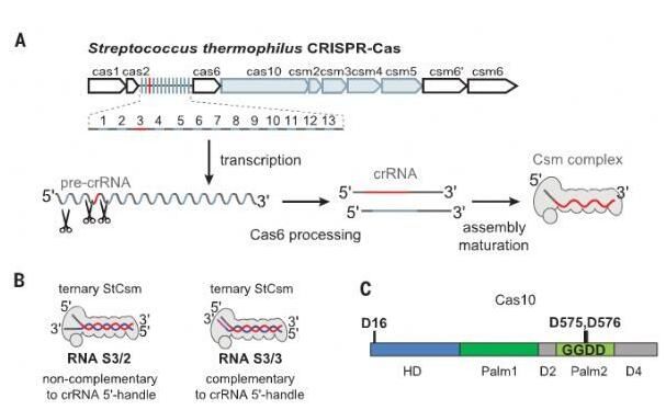
[15] Science: A major breakthrough! Revealing a circular oligoadenylation signal pathway in the type III CRISPR-Cas system
Doi:10.1126/science.aao0100 doi:10.1126/science.aao2210
In the type III CRISPR-Cas system of prokaryotes, a variety of Cas proteins are assembled with CRISPR RNA (crRNA) to form Csm (for Type III-A CRISPR-Cas systems) or Cmr (for Type III-B CRISPR-- For the Cas system, the effector complex, the Csm or Cmr complex, interferes with the invading nucleic acid by a transcription-dependent DNA silencing. After the phage infects the bacteria, its DNA begins to transcribe in order to establish and maintain the infection cycle. In bacteria, this crRNA-directed Csm/Cmr complex acts as a complementary target sequence in the RNA of the surveillance complex scanning invaders, the protospacer.
The crRNA directs Csm/Cmr to bind to the invader's RNA, triggering the Csm3/Cmr4 subunit to cleave this RNA and simultaneously activating the single-strand DNase activity of the Cas10 subunit, thereby degrading the single strand in the transcription bubble DNA. The Csm or Cmr complex avoids autoimmune responses by examining the complementarity between the 5'-handle of the crRNA and the 3' end flanking sequence of the RNA target sequence. The base pairing of the crRNA 5'-handle with the target RNA of the phage inhibits the single-strand DNase activity of Cas10, thereby protecting the host DNA from degradation. The absence of complementarity between the crRNA 5'-handle and the phage target RNA would indicate that the single-stranded DNA in the transcriptional vesicle is a non-self DNA template, thereby activating the single-strand DNase activity of Cas10 and degrading it.
The Cas10 subunit is also referred to as Csm1 in the III-A type CRISPR-Cas system and Cmr2 in the III-B type CRISPR-Cas system. It contains an N-terminal HD domain, two small alpha helix domains and two Palm domains. Both Palm domains have a ferredoxin-like fold, and this folded region has the core domain of a nucleic acid polymerase and a nucleotide cyclase.
[16] Science: a new mechanism for brain memory formation
DOI: 10.1126/science.aan6203
Using the new "NeuroGrid" technology, scientists have discovered that sleep promotes interaction between the brain and areas of memory formation. The results were published in the journal Science.
A type of structure in the brain called the hippocampus has a key role in the transformation of newly formed memory into permanent memory. Previously, researchers have discovered that during the sleep phase, the hippocampus of the brain produces a high-frequency neural signal that they believe Signals play an important role in the storage of memory. The current study demonstrates the existence of this signal and demonstrates that their specific distribution sites are in areas of the brain where complex susceptibility information is processed.
"When we first discovered it, we thought it was wrong, because this signal has never been discovered before," said Dion Khodagholy, assistant professor of Columbia University, the first author of the study.
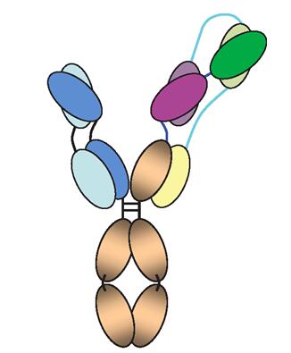
[17] Science: Heavy! A three-pronged three-specific antibody is expected to prevent HIV infection
Doi:10.1126/science.aan8630
In a new study, American researchers reported that a three-pronged antibody (trispecific antibody) made in the laboratory is better than a single natural antibody used to make this trispecific antibody. Monkeys were protected from infection by two human monkey chimeric immunodeficiency virus (SHIV) strains. The results of the study were published online September 20, 2017 in the journal Science, entitled "Trispecific broadly neutralizing HIV antibodies mediate potent SHIV protection in macaques".
This trispecific antibody was developed by researchers from the National Institutes of Health (NIH) and the French pharmaceutical company Sanofi, and it also blocks more HIV in the laboratory than a single natural antibody. The strain infects the cells. This new GF-binding antibody binds to three distinct key sites of HIV.
Currently, these researchers are planning to conduct early clinical trials of this trispecific antibody in healthy people and HIV-infected people, hoping that it will eventually be used to prevent and treat HIV for a long time. By combining with three different sites of HIV, the virus should be more difficult to evade than the single natural antibody.
[18]Science: The new method produces bryozoans that have the potential to treat cancer and HIV, yielding tens of thousands of times more productive
Doi:10.1126/science.aan7969 doi:10.1126/science.aao5346
A drug isolated from marine pests is expected to treat some of the most serious diseases, and scientists want to know how effective it is - as long as they can get more of it. As far as the current situation is concerned, the supply of this chemical in the world has fallen to about half of that in the 1990s, and it is difficult to extract enough quantities from the marine organisms that produce it.
Now, in a new study, researchers from Stanford University in the United States have found a simpler, more efficient way to make such increasingly demanding compounds in the lab. Their newly synthesized drugs will be sufficient to continue clinical trials to test its efficacy as a cancer immunotherapy and to treat Alzheimer's disease and HIV. The results of the study were published in the October 13, 2017 issue of Science, titled "Scalable synthesis of bryostatin 1 and analogs, adjuvant leads against latent HIV."
Paul Wender, the author of the paper and a professor of chemistry at Stanford University, said that he was very excited about the project. "I put on my lab coat and did some crystallization experiments." For him, this paper is the result of decades of research.
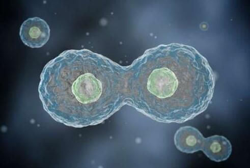
[19]Science: Using DNA replication rhythm to kill cancer cells
Doi:10.1126/science.aao3172 doi:10.1126/science.aaq0678
Human cells produce new cells through division during their lifetime. In this process, stable or even rhythmic supply of DNA building blocks is necessary to generate new DNA. Now, in a new study, researchers from the School of Health and Medicine at the University of Copenhagen in Denmark demonstrated for the first time how human cells can precisely regulate this process to ensure that it does not go wrong and cause disease. They also showed how they can manipulate this rhythm and point out how this can be used to kill cancer cells in the future. The results of the study were published in the November 10, 2017 issue of Science, titled "Redox-sensitive alteration of replisome architecture safeguards genome integrity."
In human cells, new DNA is formed using a constituent unit called a ribonucleotide reductase (RNR) called a nucleotide. Prior to this, we have not fully understood how the RNR rhythm and the presence of a suitable number of nucleotides are exactly consistent with the rate of DNA replication.
Today, these researchers draw the flow and regulation of nucleotides. This flow follows the same rhythm as DNA replication, and when deviations occur, the cell regulates the process and keeps the two consistent.
[20] Science: A major breakthrough! Building the world's smallest tape recorder with the bacterial CRISPR/Cas system
Doi:10.1126/science.aao0958
In a new study, researchers from the Columbia University Medical Center used a clever molecular hacking technique to turn a natural bacterial immune system into a tiny data logger for the development of bacterial cells. New technologies for disease diagnosis and environmental monitoring lay the foundation. The relevant research results were published online in the journal Science on November 23, 2017, and the paper titled "Multiplex recording of cellular events over time on CRISPR biological tape".
These researchers genetically modified a common laboratory strain of Escherichia coli, which is ubiquitous in the human gut, so that they not only recorded their interactions with the environment, but also recorded when these events occurred.
"These bacteria swallowed by patients may be able to record the changes they have experienced throughout the digestive tract," said Harris Wang, an assistant professor of thesis and assistant professor of pathology, cell biology and systems biology at Columbia University Medical Center. The phenomenon has reached an unprecedented level of understanding. Other applications may include basic research in environmental monitoring, ecology and microbiology. (Bio Valley Bioon.com)
Source: Sina Technology
下一条: The University of Tokyo has developed a hard polymer glass that can be self-healing and healing.
Address:Tianan Smart City A1-602, No. 228 Linghu Avenue, Xinwu District, Wuxi, Jiangsu, China TelePhone:0510-85386636 Fax:0510-85384339 E-mail:info@solmontech.com
KeyWord:

